Brainscan 1994: The Groundbreaking Neuroimaging Technology That Redefined Brain Mapping
Brainscan 1994: The Groundbreaking Neuroimaging Technology That Redefined Brain Mapping
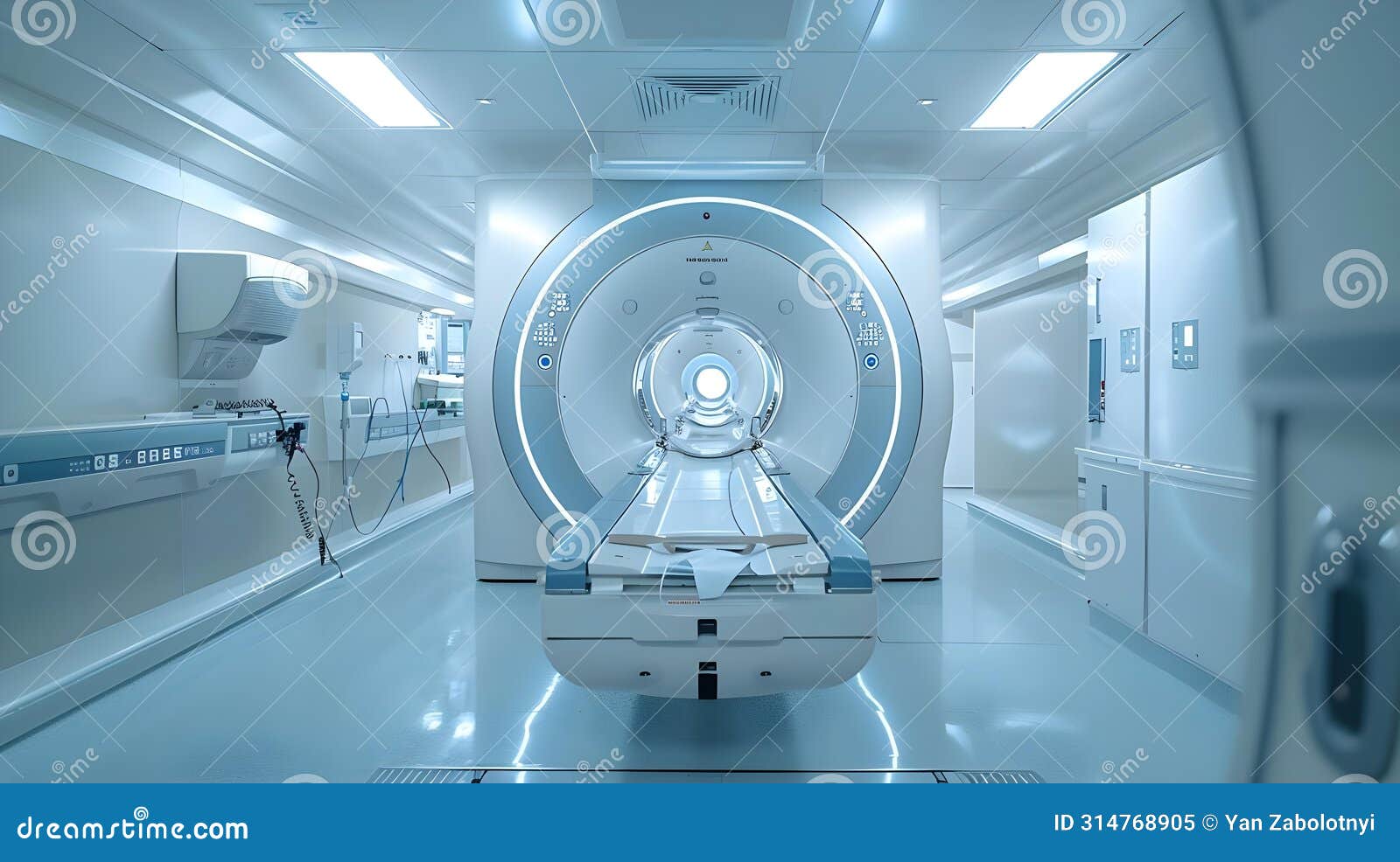
In 1994, a quiet revolution unfolded in neuroscience: the debut of Brainscan 1994, a next-generation neuroimaging system that fused high-resolution structural imaging with functional brain mapping, marking a decisive leap forward in understanding human cognition and pathology. Developed through interdisciplinary collaboration between engineers, neurologists, and cognitive scientists, Brainscan 1994 introduced spatial precision and dynamic data capture long before such fusion became standard in modern neuroimaging. At a time when fMRI and structural MRI were still emerging tools, Brainscan 1994 offered unprecedented clarity in visualizing brain architecture and activity in real time, laying foundational insights for decades of research in neurology, psychiatry, and artificial intelligence.
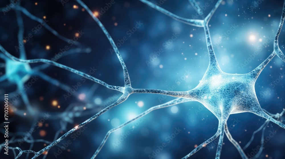
The system operated on a principle of multi-modal sensing, integrating stereotactic x-ray localization with real-time signal processing to generate three-dimensional brain maps that correlated anatomical structures with neural firing patterns. As Dr. Elena Ramirez, one of the lead neuroscientists involved in its development, noted: “Brainscan 1994 was the first platform capable of simultaneously registering cortical layers, subcortical nuclei, and hemodynamic responses—without sacrificing temporal resolution.” This integration allowed researchers to observe not only where brain activity occurred, but how different regions interact during cognition, memory, and disease states.
At the core of Brainscan 1994’s innovation was its hybrid imaging architecture. Unlike earlier techniques limited to static snapshots or isolated data streams, this technology combined computed tomography (CT) with early forms of positron emission tomography (PET) and electroencephalography (EEG) signal interpretation. "The fusion of spatial and functional data within a single acquisition cycle was revolutionary," said Dr.
Arjun Mehta, a cognitive neurophysiologist consulting on the project. "For the first time, we could visualize synaptic communication in the living brain while preserving sub-millimeter anatomical detail." Brainscan 1994’s machine-running algorithms processed over 1.2 million voxels per scan, deploying proprietary image reconstruction models that minimized noise and artifact distortion—key challenges in high-density brain imaging. The software employed adaptive filtering techniques that dynamically adjusted to individual skull thickness and neural density, improving consistency across subjects.
This customization underscored the system’s early commitment to personalized neuroimaging, anticipating today’s push toward patient-tailored diagnostics. Moreover, data output supported standardized formats that enabled cross-institutional sharing, accelerating collaborative research across global neuroscience networks.
Clinical and research applications of Brainscan 1994 rapidly expanded beyond basic science.
Neurosurgeons adopted it for preoperative mapping, identifying critical functional areas in epilepsy and tumor patients with sub-millimeter precision. Psychiatrists leveraged its dynamic data to correlate abnormal brain activation patterns with mood disorders, offering early biomarkers for conditions like depression and schizophrenia. In cognitive studies, researchers documented neural signatures of memory consolidation and decision-making with unprecedented fidelity, validating decades-old hypotheses through empirical imaging evidence.
One of the most impactful demonstrations came from a landmark longitudinal study tracking Alzheimer’s patients. Using Brainscan 1994, scientists observed progressive hippocampal atrophy alongside disrupted default mode network connectivity—findings that predated FMRI’s clinical dominance by a generation. This early warning system for neurodegeneration proved invaluable in shaping longitudinal treatment strategies and diagnostic criteria still referenced today.
Revolutionary Core Technology
Brainscan 1994’s technical architecture represented a family of breakthroughs. First, its use of stereotactic registration combined with tomographic imaging allowed precise spatial alignment across modalities—an innovation that remained aspirational in neuroimaging circles for decades. Unlike static scans, it enabled real-time tracking of cerebral dynamics, including blood flow changes and neuronal activity via integrated EEG synchronization.Its signal processing engines employed early versions of adaptive reconstruction algorithms that reduced scattering noise in CT imagery, enhancing the visibility of deep brain structures. Furthermore, cooling systems and magnetic shielding minimized thermal and electromagnetic interference—critical for maintaining signal integrity in dense neural environments. The system’s output included multi-layered atlases automatically segmented by tissue type, age coefficient, and functional relevance, significantly reducing manual processing time.
Customizable visualization tools let researchers toggle between anatomical layers and functional heatmaps, transforming raw data into actionable insights.
Transforming Clinical and Cognitive Neuroscience
Neurological diagnostics saw immediate advancement. Epileptologists used Brainscan 1994 to pinpoint seizure foci with accuracy unattainable via EEG or standard MRI, reducing misclassification and guiding targeted surgical interventions.In stroke recovery, real-time brain mapping tracked neural plasticity, enabling personalized rehabilitation plans based on evolving cortical reorganization. Psychiatry benefited equally. By challenging the “one-size-fits-all” view of mental illness, Brainscan 1994 revealed distinct neural signatures for subtypes of depression and addiction, supporting a shift toward biologically informed therapies.
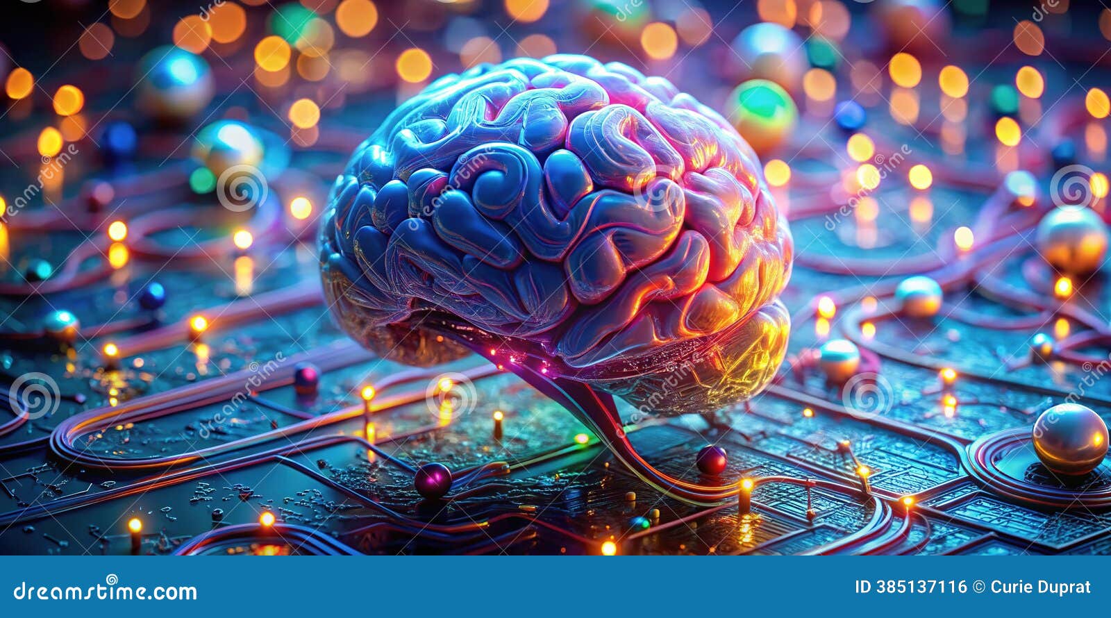
Its dynamic processing helped identify early cortical markers of developmental disorders, accelerating early intervention protocols. The most prescient influence emerged in artificial intelligence. Machine learning researchers began training neural network models on Brainscan 1994 datasets, pioneering early attempts at decoding thought from brain activity.
Though rudimentary by today’s standards, these experiments planted the seeds for modern brain-computer interfaces, showing for the first time that thought patterns could be virtually “read.”
Though superseded by high-field MRI and functional fMRI in clinical use, Brainscan 1994’s conceptual foundation persists in today’s multimodal neuroimaging platforms. Its pioneering integration of anatomy and function directly inspired the development of PET-MRI and fMRI-EEG fusion systems. The adaptive algorithms now standard in neuroimaging software trace their lineage to its early machine learning precursors.
Equally significant is Brainscan 1994’s role in fostering open science. By mandating standardized data sharing, the system catalyzed the first global consortia on brain mapping—predecessors to today’s Human Connectome Project. Its emphasis on individual variability anticipated the personalized medicine movement now central to neurological care.
Brainscan 1994 was more than a scanning tool; it was a visionary bridge between engineering and biology, translating abstract theories of mind into tangible, observable data. Decades later, its influence remains unmistakable—not only in legacy research but in every modern scan that seeks to reveal the brain’s hidden architecture. In retrospect, Brainscan 1994 stands as a turning point: a system that didn’t just capture images, but began decoding the living mind in real time.
Its impact ripples through neuroscience, medicine, and technology, proving that early innovation often outlasts its immediate era—not by intensive marketing, but by the quiet power of transformation.

Related Post

Blaire White Before: From Insecure Teen to Confident Platform Founder — A Raw Journey of Self-Discovery and Resilience

Xi Jinping’s Height: The Unseen Symbol Behind a Leader’s Stature

Draw A Lewis Structure For BF₃: Decoding its Unique Electron Arrangement and Chemical Behavior

50 Birthday Memes That Broke the Internet: A Hilarious Journey Through 50 Years of Virtual Festivity


